
Digital Dental X-Ray in Pathirippala
Precise, low-dose digital dental imaging in Pathirippala, Palakkad. At Complete Dental Clinic, diagnostic radiographs help our dentists examine tooth roots, bone support, and sub-surface areas that cannot be fully evaluated through visual examination alone, ensuring safe and accurate treatment planning.
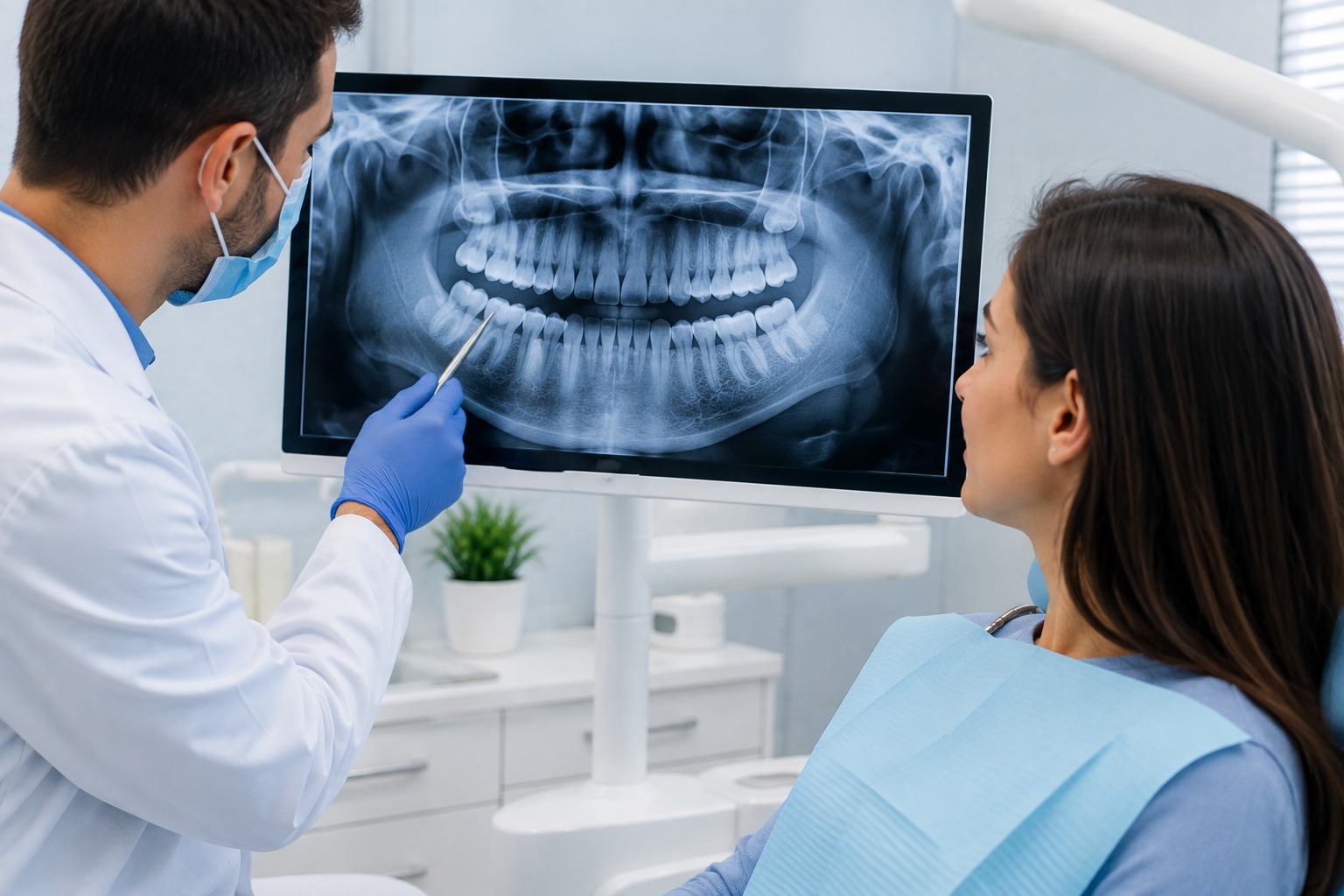
Understanding Digital Dental Radiography
A routine dental checkup allows a dentist to inspect the visible enamel surfaces, biting ridges, and gum margins inside your mouth. However, substantial portions of each tooth—including the internal dentin, root canals, and surrounding jawbone—lie beneath the gumline and cannot be fully evaluated through visual examination alone.
Digital dental radiography utilizes electronic sensors to capture detailed dental images directly into a computer system. Instead of using traditional chemical film, digital technology allows immediate high-resolution viewing on chairside monitors, supporting diagnostic precision while minimizing radiation exposure.
Sub-Surface Clarity
Visualizing tight interproximal contact zones, root tips, and supporting bone margins hidden from ordinary view.
Immediate Chairside Review
Digital images appear instantly on the clinic monitor, eliminating darkroom processing delays.
Targeted Patient Safety
Carefully prescribed under ALARA principles, ensuring imaging occurs only when clinically beneficial.
Why Might a Dental X-Ray Be Recommended?
Dentists recommend diagnostic imaging when specific symptoms, clinical findings, or treatment requirements necessitate an in-depth look inside teeth or bone structures:
Interproximal Tooth Decay
Cavities forming between tightly contacting teeth are shielded by enamel and often cannot be detected early without imaging.
Recurrent Decay Under Restorations
Demineralization or decay developing beneath existing tooth fillings, inlays, or dental crowns along marginal edges.
Periodontal Bone Support Levels
Evaluating the crestal bone height surrounding tooth roots to diagnose gum disease progression or monitor stability.
Root Infections & Periapical Findings
Revealing bone changes suggestive of periapical inflammation, infection, or pathology around root tips, interpreted alongside clinical symptoms and vitality testing.
Impacted & Unerupted Teeth
Assessing the position, angle, and root curvature of impacted wisdom teeth or unerupted adult canines prior to removal.
Dental Trauma & Root Fractures
Evaluating cracked teeth, horizontal root fractures, or jawbone damage following accidental blows or sports impacts.
Types of Dental Radiographs Used in Clinical Practice
Different clinical scenarios require specific imaging techniques depending on whether the dentist needs to evaluate a single tooth or obtain a broader view of the facial bones:
Bitewing Radiographs
Bitewing X-rays capture the crowns of both upper and lower back teeth in a single frame as you bite down on a sensor tab.
- Detects early decay between contacting posterior molars.
- Evaluates bone height between adjacent teeth for gum stability.
Periapical (IOPA) Radiographs
Intraoral Periapical images show the entire tooth from the chewing crown down past the root tip into the jawbone.
- Essential for evaluating root canal anatomy and infections.
- May reveal findings suggestive of periapical lesions, root fractures, or bone changes.
Panoramic (OPG) Imaging
An extraoral panoramic radiograph that rotates around the head to capture a wide continuous view of both upper and lower jaws.
- Shows wisdom tooth positioning, impaction angles, and sinuses.
- Assesses temporomandibular joint (TMJ) bony architecture.
CBCT / 3D Volumetric Imaging
Cone Beam Computed Tomography produces three-dimensional cross-sectional slices of bone volume and anatomical structures.
- Used when detailed 3D depth is needed for dental implants.
- Evaluates nerve canal relationships before surgical extractions.
Digital Imaging vs. Conventional Film
Digital radiography represents a substantial technological advancement over legacy film-based X-rays, delivering practical benefits for patient safety and clinical diagnosis:
Reduced Radiation Exposure
Digital sensors are significantly more sensitive to X-ray beams than photographic film, allowing clear images to be acquired with substantially lower radiation exposure.
Instantaneous Image Preview
Images appear on the computer monitor within seconds of exposure, eliminating chemical darkroom processing time and shortening appointment durations.
Image Zoom & Contrast Enhancement
Dentists can magnify small areas, adjust brightness, and highlight subtle density changes, allowing you to clearly see and understand your dental health findings.
Environmentally Responsible
Because electronic files replace physical film, the process eliminates chemical developers, fixers, and toxic lead foil disposal from the clinical environment.
Radiation Safety & The ALARA Principle
While dental X-rays involve ionizing radiation, the dosage associated with modern digital dental imaging is remarkably low. Every day, people naturally absorb small amounts of background radiation from cosmic sources, soil, rocks, and everyday surroundings. A standard set of digital dental X-rays contributes only a tiny fraction of that routine background exposure.
The ALARA Principle: Dental professionals adhere strictly to the international safety standard known as ALARA—As Low As Reasonably Achievable. This means imaging is never performed routinely without purpose; each scan must be clinically justified to provide meaningful diagnostic benefit that aids your dental health.
Pregnancy Considerations: If you are pregnant or suspect you might be, inform your dental team before any procedure. When diagnostic imaging is clinically necessary to evaluate pain, infection, or acute conditions, dental radiographs can be considered following individual clinical evaluation and appropriate radiation-safety precautions, while non-essential imaging may be deferred.
What to Expect During Your Dental X-Ray
Dental imaging is a quick and non-invasive procedure. While the X-ray exposure itself is completely painless, holding an intraoral sensor in position may feel briefly uncomfortable for some patients. A standard intraoral imaging session follows five simple steps:
Clinical Need Assessment
Your dentist examines your oral cavity and determines whether specific radiographic imaging is clinically warranted for diagnosis.
Comfortable Positioning
You are seated comfortably in the dental chair, and appropriate clinical protective protocols are put in place.
Sensor Placement
A small digital sensor enclosed in a sterile, single-use barrier sleeve is gently positioned next to the tooth, and you rest your teeth together on a bite guide.
Brief Exposure
The imaging arm is aligned outside your cheek. You remain still for just a fraction of a second while the low-dose exposure occurs.
Instant Collaborative Review
The image appears immediately on the screen, allowing you and your dentist to review the findings and discuss your care plan together.
How Dental Imaging Supports Specialized Treatments
Digital dental X-rays provide essential diagnostic mapping for restorative, surgical, and preventive dental therapies:
Root Canal Treatment
Identifies canal numbers, curvature, root lengths, and monitors periapical healing.
Dental Implants
Evaluates jawbone thickness, density, sinus floors, and critical nerve canal proximity.
Tooth Extraction
Assesses curved root morphology and surrounding bone before simple or surgical removal.
Braces & Orthodontics
Maps jaw growth relationships, tooth angulations, and unerupted permanent dentition.
Pediatric Dental Radiography Considerations
Children's oral anatomy develops rapidly as baby teeth mature and permanent teeth begin to emerge. Because children are more biologically sensitive to radiation than adults, pediatric dental radiography is conducted with strict clinical prudence.
When are X-rays needed for children? Dentists recommend pediatric imaging only when visual examination leaves unanswered diagnostic questions. Common reasons include checking for hidden cavities between contacting primary molars, monitoring the eruption pathway of adult teeth, identifying congenitally missing teeth, or evaluating tooth trauma from play or sports.
When imaging is clinically indicated for children, exposure settings are carefully adjusted to account for a child's smaller anatomical size. Learn more about our dedicated pediatric dentistry services.
Clinical Safeguards & Diagnostic Limitations
While digital dental X-rays are an invaluable diagnostic asset, they are one component of a comprehensive oral examination rather than a stand-alone diagnostic test.
Two-Dimensional Projection
Standard radiographs compress three-dimensional anatomical structures into flat images. Overlapping tooth surfaces can sometimes mask subtle, very early enamel changes.
Soft Tissue Limitations
X-rays pass easily through soft oral tissues. While they reveal hard bone density and mineralized tooth enamel, they cannot directly diagnose soft tissue lesions or gum ulcers.
For these reasons, radiographic findings are always interpreted in combination with tactile dental probing, visual clinical assessment, periodontal charting, and your medical history.
When Should You Seek a Diagnostic Dental Assessment?
You should arrange a professional dental examination if you experience any of the following symptoms:
Digital Dental X-Ray in Pathirippala, Palakkad
Complete Dental Clinic provides comprehensive general dental care and digital diagnostic imaging in Pathirippala. Our clinical approach emphasizes patient comfort, clear diagnostic explanations, and conservative treatment planning for individuals and families throughout Palakkad district.
Conveniently situated at 1st Floor, Maimoon Square, opposite South Indian Bank, Pathirippala, Kerala 678642, our clinic welcomes patients from Pathirippala, Mankara, Parali, Ottapalam, and neighbouring communities seeking thorough diagnostic examinations, pain relief assessments, and restorative dental care.
Related Dental Services at Complete Dental Clinic
Digital radiography connects naturally with all areas of restorative and preventive dentistry:
General Dentistry
Routine oral checkups, dental cleanings, and tooth-coloured fillings for healthy smiles.
Root Canal Treatment
Targeted therapy to treat deep nerve infections and preserve natural teeth.
Tooth Extraction
Gentle, planned removal of non-restorable or problematic impacted teeth.
Pediatric Dentistry
Child-friendly checkups, cavity prevention, and eruption monitoring for growing smiles.
Frequently Asked Questions About Dental X-Rays
Helpful, clinically accurate answers to common questions regarding dental radiography:
Are dental X-rays safe?
Modern digital dental X-rays utilize highly sensitive electronic sensors that require minimal radiation exposure compared to older traditional film techniques. Dentists follow the ALARA principle (As Low As Reasonably Achievable), recommending imaging only when there is a clear diagnostic need to assess tooth roots, bone support, or hidden decay.
How often do I need dental X-rays?
Dental X-rays are not needed at every routine visit. The frequency depends on your individual oral health, symptoms, risk of tooth decay or gum disease, and ongoing dental treatments. Your dentist evaluates your clinical condition before recommending any diagnostic imaging.
Do dental X-rays hurt?
The X-ray exposure itself is completely painless and produces no physical sensation. For intraoral images, a digital sensor is placed briefly in the mouth while you bite gently on a holder. While the sensor edges can feel firm or slightly uncomfortable against the gums or palate for a few seconds, the procedure is fast and non-invasive.
Are X-rays necessary before root canal treatment?
Radiographs are an important part of root canal assessment and treatment. They assist dentists in evaluating tooth roots, surrounding bone, and signs suggestive of periapical infection, and help monitor treatment progress, though complex or fine canal anatomy may require additional clinical assessment.
Are dental X-rays safe during pregnancy?
If you are pregnant or may be pregnant, inform your dentist before any examination. Clinically necessary dental radiographs can be considered when needed to evaluate pain, infection, or urgent dental issues, using appropriate radiation-safety precautions based on individual clinical evaluation, while non-essential imaging may be deferred.
Do children need dental X-rays?
Children may occasionally require dental X-rays to check for hidden decay between tightly spaced baby teeth, monitor jaw development, or evaluate whether permanent teeth are erupting normally. Because developing tissues are sensitive, imaging for children is strictly individualized and conducted with low-dose settings only when clinically necessary.
What is the difference between OPG and IOPA?
An IOPA (Intraoral Periapical) X-ray focuses closely on one or two specific teeth from crown to root tip, offering fine detail for cavities, root infections, and bone margins. An OPG (Orthopantomogram) is an extraoral panoramic scan showing the entire upper and lower jaw, sinuses, and jaw joints in a single broad view.
Is CBCT required for dental implants?
Cone Beam Computed Tomography (CBCT) provides three-dimensional cross-sectional imaging of the jawbone. While standard two-dimensional X-rays are sufficient for many assessments, 3D imaging may be recommended for complex dental implant planning, nerve tracing, or surgical extractions when detailed anatomical depth is clinically necessary.
Need a Dental Examination or X-Ray?
If you have tooth pain, gum swelling, or require diagnostic evaluation for a treatment plan, contact Complete Dental Clinic in Pathirippala to schedule a clinical consultation.